


It is caused by herpes viruses, including herpes simplex virus (HSV), varicella zoster virus (VZV) and cytomegalovirus (CMV).
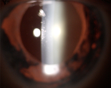
The disease can be acute and recurrent or chronic, typically unilateral and associated with elevated intraocular pressure, loss of iris tissue (iris atrophy), and deposits on the back of the cornea (keratic precipitates).
However, each viral type is paired with tell-tale features and epidemiologic clues that can help the diagnosis.
HSV and VZV associated anterior uveitis can affect people of any age.
However, HSV anterior uveitis tends to be more common in patients under 50 years old, whereas anterior uveitis related to VZV is more frequent among older patients after 60 years old.
HSV and VZV anterior uveitis are often associated with acute high intraocular pressure, keratic precipitates on the posterior surface of the cornea and iris atrophy, sometimes resulting in persistent pupillary dilatation.
Superficial corneal involvement is usually not concomitant with the uveitis but can be present in form of the so called ‘dendritic ulcer’ or inflammation of the centre of the cornea (stroma keratitis).
Severe disease can result in decreased corneal sensation and corneal scars affecting the quality of vision.
Herpetic disease can be associated with skin involvement. Patients with ocular HSV can give a history of cold sores affecting the lips or genital ulcers, , usually not concomitant with an episode of uveitis.
VZV is the cause of both childhood chicken pox and, as reactivated infection, shingles later in life.
The reactivation is caused by the virus which re-activates from its dormant status in the cells of the central nervous system, and travels along neurons to the the skin to manifest the disease often referred to as herpes zoster.
Herpes zoster ophthalmicus occurs when the virus reactivates in the areas of the skin innervated by trigeminal nerve around the eye.
The disease is characterized by a unilateral, painful, erythematous, skin rash with vesicles, pustules and crusting lesions. Uveitis usually follows the skin eruption.
CMV uveitis can affect patients at every age and tends to be more common in Asian population and in males.
The clinical presentation of uveitis with high ocular pressure and other typical ocular features is sufficient to raise the suspicion of a viral diagnosis (usually straightforward in the setting of skin involvement).
However, more challenging than diagnosing herpetic anterior uveitis can sometime be distinguishing which herpes virus is the cause of the inflammation.
A confirmed diagnosis can be obtained with a diagnostic sampling of intraocular fluid taken and analysed for the detection of the virus.
Topical and systemic antiviral medications, including acyclovir and valacyclovir are the mainstay of therapy in the management of HSV and VZV diseases, whereas ganciclovir and valganciclovir are used in the treatment of CMV related uveitis.
Antiviral medications are administered both for the treatment of acute disease as well as long- term prophylaxis to prevent recurrences.
In addition to antiviral therapy, corticosteroid eye drops are used to reduce the inflammation, and lowering pressure eye drops are administered to control intraocular pressure.
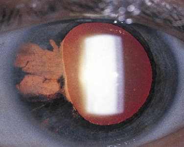
Herpetic anterior uveitis with loss of iris tissue (iris atrophy) seen as transillumination defect of the iris.

Herpetic anterior uveitis with loss of iris tissue (iris atrophy) and deposits on the back of the cornea (keratic precipitates).

Contact
Ilaria Testi
The London Clinic Eye Centre
119 Harley Street
London W1G 6AU
COPYRIGHT 2026 | Dr ILARIA TESTI | Cookie Policy | Term and Conditions