


Fluorescein angiography provides information on the retina and blood vessels of the eye, enabling the eye specialist to determine whether treatment is needed.
In particular, it is useful in showing leaking blood vessels and/or highlighting areas of the eye where the blood supply is poor.
Indocyanine green angiography provides information about the choroid, which can be affected by an inflammatory process.
Both tests are commonly used to help in the diagnosis of a particular disease, to assess it severity, and as a monitoring to assess the response to treatment and the presence of complications.
The procedure has been carried on for several decades and has been proven safe to use in both younger and older patients.
Patients will be asked questions regarding their general health, medications and any allergies they may have, including to a previous administration of dye.
A consent form needs to be signed before the procedure, to confirm the patient has agreed to the treatment and that understands the benefits and risks.
Dilating drops will be administered to enlarge the pupils.
The procedure involves putting a small plastic tube into a vein of the arm or hand (intravenous cannula), through which the dye will be administered. The dye will then be injected and within seconds will reach the blood vessels in the eyes. Patients will be sitting still in front of a camera with the chin on the chin rest and the head firmly forward against the band.
A series of photos of the back of each eye will be taken in quick succession. Patients will be asked to sit back for a few minutes before a further series of photos will be taken.
Patients will be asked to stay in the clinic for at least half an hour after administering the dye to observe any adverse reaction to it. The cannula will then be removed before going home.
After the injection of the dye, skin and urine will turn yellow for approximately 24–48 hours. This is normal and not harmful.
Patients will be advised to drink plenty of water so to help flush the dye through the body.
Nausea might be experienced when the dye is flushed through. This usually passes within seconds and is helped by deep breathing.
Other side effects include dizziness, vomiting, itchy skin and/or skin rash, but these are not as common.
More serious, but very rare, adverse reactions include bronchospasm (constriction of the airway), shortness of breath and anaphylactic shock, which may lead to collapse.
Adverse reactions would receive immediate attention by the hospital team. If patients experience shortness of breath or feel faint or generally unwell, they should inform the team immediately and appropriate care will be given.
In rare cases the dye may leak outside the vein into the skin causing pain or burning sensation. This can be relieved by placing ice packs to the affected area.
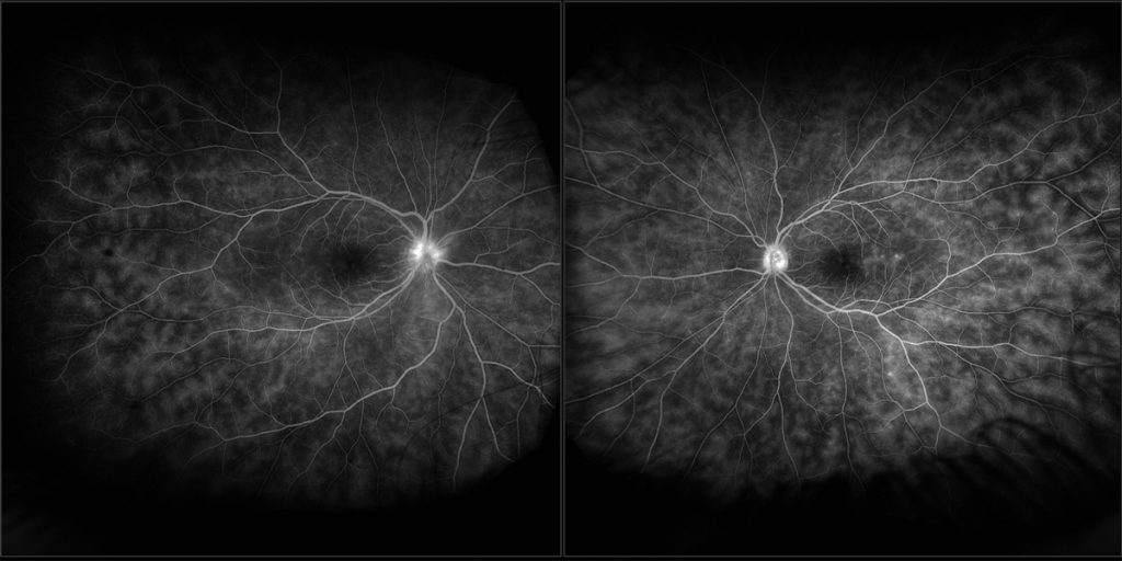
Fluorescein angiography images showing bilateral retinal vasculitis in Behçet’s disease.

Fluorescein angiography images showing peripheral areas of vascular non-perfusion with retinal neovascularization in patient with tubercular occlusive vasculitis.
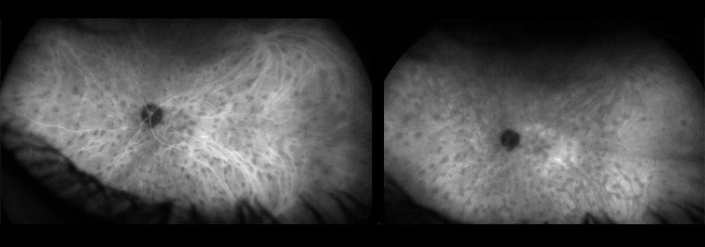
Indocyanine green angiography images showing active choroidal inflammation (dark dots) In a patient with Birdshot retinochoroiditis.

Contact
Ilaria Testi
The London Clinic Eye Centre
119 Harley Street
London W1G 6AU
COPYRIGHT 2026 | Dr ILARIA TESTI | Cookie Policy | Term and Conditions