


The disease usually manifests a prodromal phase characterized by flu like symptoms, including fever, headache and neck stiffness.
There can be dysacusia (discomfort caused by loud noises or distortion in the quality of the heard sounds), tinnitus (perception of sound with no corresponding external sound often described as a ringing), and dizziness (vertigo). However, some patients report no symptoms.
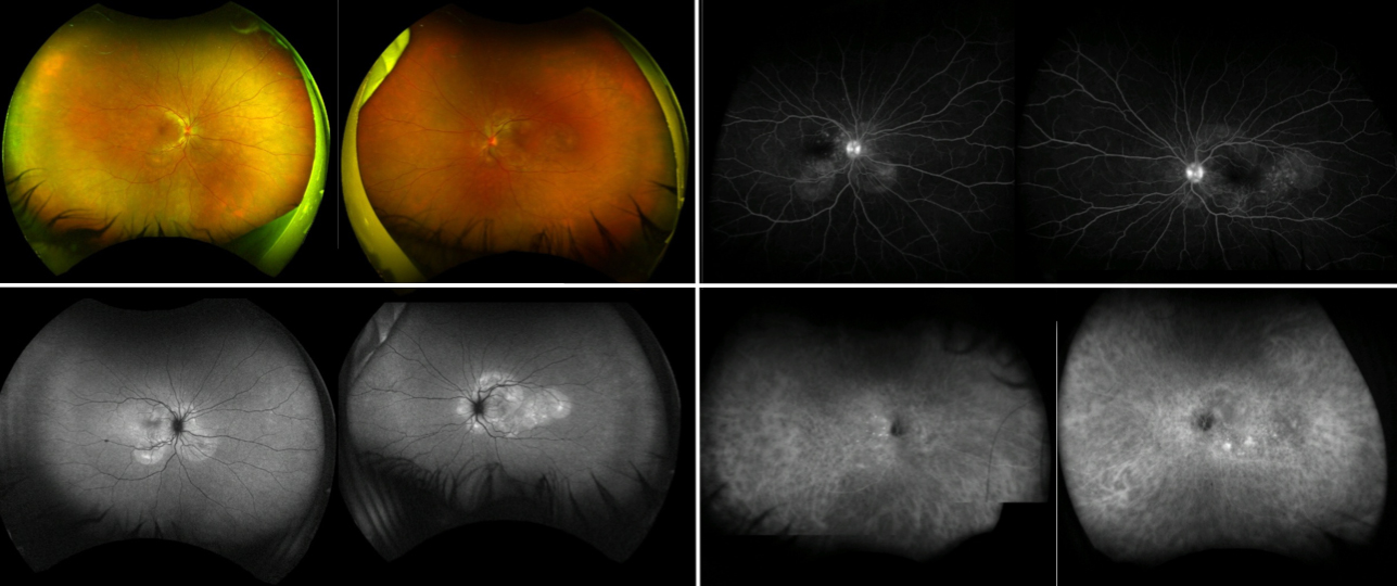
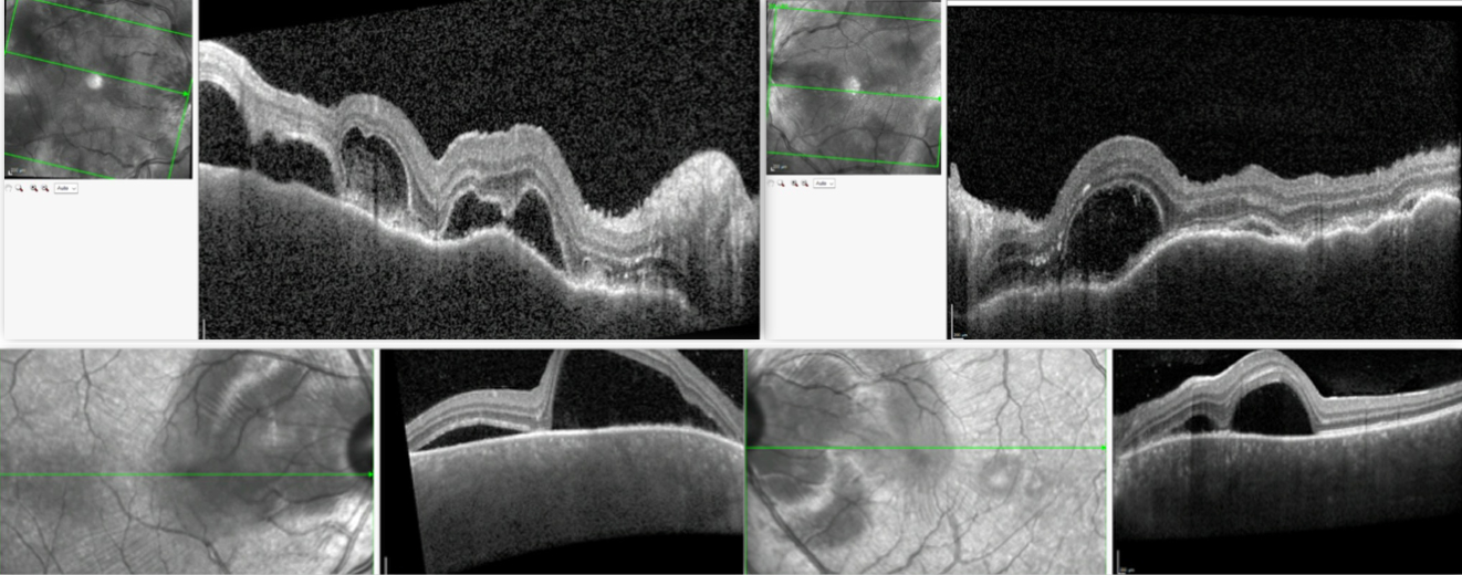
The acute phase of the ocular disease occurs a few days after the prodromal phase and manifests as a bilateral uveitis, typically characterized by massive accumulation of fluid below the retina causing multiple exudative bullous retinal detachments, associated with intraocular inflammation and optic nerve inflammation.
The chronic phase after the acute stage is characterized by changes in the skin, including loss of colour of the hair and eyelashes (poliosis), of the skin (vitiligo) and hair loss (alopecia), and depigmentation of the tissues at the back of the eye, including the choroid, resulting in a classic orange-red discoloration at the back of the eye called “sunset glow fundus”.
Recurrences of the disease most commonly manifest as bilateral anterior uveitis, but may also affect the back of the eye.
VKH is an inflammatory condition presumably driven by an abnormal immune response against the melanocytes occurring in genetically predisposed individuals.
Melanocytes are specialized skin cells that produce a protective skin-darkening pigment called melanin and that can be found in the eyes, ear, skin and central nervous system (meninges).
The disease cannot be passed on to other people or from parent to child, but genetic factors may increase the likelihood of a person developing the condition.
The diagnosis of VKH is based on the classic systemic and ocular features after excluding other diagnosis potentially causing similar ocular manifestations. There are no specific laboratory tests to diagnose VKH.
Optical coherence tomography is an ocular imaging technique that is performed in patients with clinical suspicion of VKH to detect exudative retinal detachments.
Fluorescein angiography is helpful in demonstrating the leakage which results in the fluid accumulation while Indocyanine angiography can be performed to detect choroidal inflammation, to monitor the course of the disease and its response to treatment. The retinal function can be measured by special tests, including visual fields and electrodiagnostic tests.
The intention of therapy is to control the inflammation and prevent the damage to the retina and the choroid, and consequently the visual loss.
The treatments are not curative but aim at controlling the immune response which is the reason for the inflammation that damages the ocular tissues.
Options include the use of oral medications, usually starting with systemic steroids to induce rapid response. In view of the nature of the disease, the addition of immunosuppressive drugs is important to allow long term control and minimize the side effects of systemic steroids. The early diagnosis and initiation of aggressive therapy are important for a good outcome.
If not well controlled, VKH can result in long term damage because of the chronic inflammation.
Delays or insufficient therapy tend to result in progressive damage of the retina and visual loss.
Other common complications include glaucoma, cataract and optic atrophy.
All these complications can affect the visual function.

Send us a message and we will get back to you.

Contact
Ilaria Testi
The London Clinic Eye Centre
119 Harley Street
London W1G 6AU
COPYRIGHT 2026 | Dr ILARIA TESTI | Cookie Policy | Term and Conditions